Biosynthesis of Fe1.83Mn0.5Zn0.41O4 nanoparticles from milk thistle seed extract for use as anticancer agents
DOI:
https://doi.org/10.69923/kezfk969Keywords:
Fe1.83Mn0.5Zn0.41O4, Nanoparticles, Milk Thistle, HL-60 cell line, leukaemia cancerAbstract
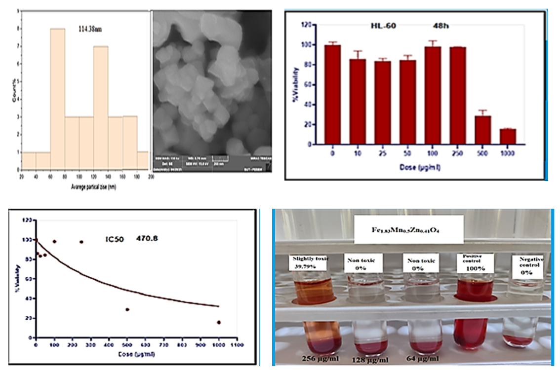
This study employed a novel, simple, and cost-effective biosynthesis approach that combined co-precipitation, ultrasonic treatment, and green chemistry techniques to synthesize Fe₁.₈₃Mn₀.₅Zn₀.₄₁O₄ nanoparticles using milk thistle (Silybum marianum) seed extract. The synthesized nanoparticles were characterized using Fourier Transform Infrared Spectroscopy (FT-IR), X-ray Diffraction (XRD), Energy-Dispersive X-ray Spectroscopy (EDX), Zeta Potential analysis, and Scanning Electron Microscopy (SEM). XRD analysis revealed an average crystallite size of 18.66 nm, whereas SEM analysis indicated an average particle size of 114.38 nm, suggesting particle aggregation. The anticancer activity of the synthesized Fe₁.₈₃Mn₀.₅Zn₀.₄₁O₄ nanoparticles was evaluated against human leukemia HL-60 cell lines. The results demonstrated significant cytotoxic activity at higher nanoparticle concentrations, with a pronounced reduction in cell viability observed at concentrations of 500 and 1000 μg/mL. The percentages of viable cells at nanoparticle concentrations of 10, 25, 50, 100, 250, 500, and 1000 μg/mL were 85.98%, 83.56%, 84.54%, 98.41%, 97.87%, 29.04%, and 15.81%, respectively. The calculated IC₅₀ value was 470.8 μg/mL. Statistical analysis using one-way ANOVA revealed highly significant differences among the treatment groups (P < 0.0001). The coefficient of determination (R² = 0.9856) indicated excellent model fitting and confirmed the strong relationship between nanoparticle concentration and biological response. The biocompatibility of the synthesized nanoparticles was further evaluated using a hemolysis assay with red blood cells at concentrations of 64, 128, and 256 μg/mL. The results showed no significant hemolytic activity or cytotoxic effects on erythrocytes, indicating that the synthesized nanoparticles were hemocompatible and safe within the tested concentration range.
Downloads
References
[1]. J. Jeevanandam, A. Barhoum, Y. S. Chan, A. Dufresne, and M. K. Danquah, “Review on Nanoparticles and Nanostructured materials: history, sources, Toxicity and Regulations,” Beilstein Journal of Nanotechnology, vol. 9, no. 1, pp. 1050–1074, Apr. 2018, doi: https://doi.org/10.3762/bjnano.9.98.
[2]. H. R. Khan, Meena Kishore Sakharkar, A. Nayak, U. Kishore, and A. Khan, “Nanoparticles for biomedical applications: An overview,” Elsevier eBooks, pp. 357–384, Jan. 2018, doi: https://doi.org/10.1016/b978-0-08-100716-7.00014-3.
[3]. M. Latha, M. Priyanka, P. Rajasekar, R. Manikandan, and N. M. Prabhu, “Biocompatibility and antibacterial activity of the Adathoda vasica Linn extract mediated silver nanoparticles,” Microbial Pathogenesis, vol. 93, pp. 88–94, Apr. 2016, doi: https://doi.org/10.1016/j.micpath.2016.01.013.
[4]. H. Barabadi, M. Ovais, Z. K. Shinwari, and M. Saravanan, “Anti-cancer green bionanomaterials: present status and future prospects,” Green Chemistry Letters and Reviews, vol. 10, no. 4, pp. 285–314, Oct. 2017, doi: https://doi.org/10.1080/17518253.2017.1385856.
[5]. R. Abbas et al., “Silver Nanoparticles: Synthesis, Structure, Properties and Applications,” Nanomaterials, vol. 14, no. 17, p. 1425, 2024, doi: https://doi.org/10.3390/nano14171425.
[6]. A. F. Al-Alawy, E. E. Al-Abodi, and R. M. Kadhim, “Synthesis and Characterization of Magnetic Iron Oxide Nanoparticles by Co-Precipitation Method at Different Conditions,” Journal of Engineering, vol. 24, no. 10, p. 60, Oct. 2018, doi: https://doi.org/10.31026/j.eng.2018.10.05.
[7]. M Haneefa, M Jayandran, and V. Balasubramanian, Asian Journal of Pharmaceutics, vol. 11, no. 1, Jan. 2017, doi: https://doi.org/10.22377/ajp.v11i01.1045.
[8]. N. E.-A. El-Naggar, M. H. Hussein, and A. A. El-Sawah, “Phycobiliprotein-mediated synthesis of biogenic silver nanoparticles, characterization, in vitro and in vivo assessment of anticancer activities,” Scientific Reports, vol. 8, no. 1, Jun. 2018, doi: https://doi.org/10.1038/s41598-018-27276-6.
[9]. S. B. Singh et al., “Structural, thermal and magnetic studies of MgxZn1−xFe2O4 nanoferrites: Study of exchange interactions on magnetic anisotropy,” Ceramics International, vol. 42, no. 16, pp. 19179–19186, Dec. 2016, doi: https://doi.org/10.1016/j.ceramint.2016.09.081.
[10]. “Smit, J. and Wijn, H.P.J. (1959) Ferrites. Philips Technical Library, Eindhoven, 150. - References - Scientific Research Publishing,” www.scirp.org. https://www.scirp.org/reference/referencespapers?referenceid=1266992
[11]. M. M. Cruz et al., “Enhanced magnetic hyperthermia of CoFe2O4 and MnFe2O4 nanoparticles,” Journal of Alloys and Compounds, vol. 703, pp. 370–380, May 2017, doi: https://doi.org/10.1016/j.jallcom.2017.01.297.
[12]. A. Amirabadizadeh, H. Farsi, M. Dehghani, and H. Arabi, “Effect of Substitutions of Zn for Mn on Size and Magnetic Properties of Mn–Zn Ferrite Nanoparticles,” Journal of Superconductivity and Novel Magnetism, vol. 25, no. 8, pp. 2763–2765, Aug. 2011, doi: https://doi.org/10.1007/s10948-011-1259-5.
[13]. A. Angermann, J. Töpfer, K. L. da Silva, and K. D. Becker, “Nanocrystalline Mn–Zn ferrites from mixed oxalates: Synthesis, stability and magnetic properties,” Journal of Alloys and Compounds, vol. 508, no. 2, pp. 433–439, Sep. 2010, doi: https://doi.org/10.1016/j.jallcom.2010.08.083.
[14]. N. T. K. Thanh and L. A. W. Green, “Functionalisation of nanoparticles for biomedical applications,” Nano Today, vol. 5, no. 3, pp. 213–230, Jun. 2010, doi: https://doi.org/10.1016/j.nantod.2010.05.003.
[15]. K. E. Sapsford et al., “Functionalizing Nanoparticles with Biological Molecules: Developing Chemistries that Facilitate Nanotechnology,” Chemical Reviews, vol. 113, no. 3, pp. 1904–2074, Feb. 2013, doi: https://doi.org/10.1021/cr300143v.
[16]. G. Nam, B. Purushothaman, S. Rangasamy, and J. M. Song, “Investigating the versatility of multifunctional silver nanoparticles: preparation and inspection of their potential as wound treatment agents,” International Nano Letters, vol. 6, no. 1, pp. 51–63, Nov. 2015, doi: https://doi.org/10.1007/s40089-015-0168-1.
[17]. S. Ahmed, M. Ahmad, B. L. Swami, and S. Ikram, “A review on plants extract mediated synthesis of silver nanoparticles for antimicrobial applications: A green expertise,” Journal of Advanced Research, vol. 7, no. 1, pp. 17–28, Jan. 2016, doi: https://doi.org/10.1016/j.jare.2015.02.007.
[18]. Philippe Lettéron et al., “Mechanism for the protective effects of silymarin against carbon tetrachloride-induced lipid peroxidation and hepatotoxicity in mice,” vol. 39, no. 12, pp. 2027–2034, Jun. 1990, doi: https://doi.org/10.1016/0006-2952(90)90625-u.
[19]. R. Saller, R. Meier, and R. Brignoli, “The Use of Silymarin in the Treatment of Liver Diseases,” Drugs, vol. 61, no. 14, pp. 2035–2063, 2001, doi: https://doi.org/10.2165/00003495-200161140-00003.
[20]. N. Vargas-Mendoza, “Hepatoprotective effect of silymarin,” World Journal of Hepatology, vol. 6, no. 3, p. 144, 2014, doi: https://doi.org/10.4254/wjh.v6.i3.144.
[21]. R. G. Mehta and R. C. Moon, “Characterization of effective chemopreventive agents in mammary gland in vitro using an initiation-promotion protocol,” Anticancer research, vol. 11, no. 2, pp. 593–6, 1991, Available: https://pubmed.ncbi.nlm.nih.gov/1905902/
[22]. Y. C. Rui, “Advances in pharmacological studies of silymarin,” Memorias do Instituto Oswaldo Cruz, vol. 86 Suppl 2, pp. 79–85, 1991, doi: https://doi.org/10.1590/s0074-02761991000600020.
[23]. Areej Ali Jarullah, Mustafa Hammadi, and K. H. Hassan, “Green Synthesis of Nano Zn3(PO4)2 Using Curcuma Extract and Their Effect on SK-OV-3 Human Ovarian Cancer Cell Lines: Effects of Nano Zn3(PO4)2 at Human Ovarian Cancer Cell,” Biological Sciences - PJSIR, vol. 68, no. 2, pp. 166–171, 2025, Accessed: Aug. 31, 2025. DOI: 10.52763/PJSIR.BIOL.SCI.68.2.2025.166.171.
[24]. H. S. Ahmed, M. Hammadi, and W. Majeed, “Green Synthesis of CuCr2O4Nano Composite from Rosemary Extract and Evaluation of its Anti-Breast Cancer Properties,” Journal of Nanostructures, vol. 14, no. 4, pp. 1183–1190, Oct. 2024, doi: https://doi.org/10.22052/JNS.2024.04.018.
[25]. M. Pagano and C. Faggio, “The use of erythrocyte fragility to assess xenobiotic cytotoxicity,” Cell biochemistry and function, vol. 33, no. 6, pp. 351–355, Aug. 2015, doi: https://doi.org/10.1002/cbf.3135.
[26]. R. D. Waldron, “Infrared Spectra of Ferrites,” Physical Review, vol. 99, no. 6, pp. 1727–1735, Sep. 1955, doi: https://doi.org/10.1103/physrev.99.1727.
[27]. Ch. S. Lakshmi, Ch. S. L. N. Sridhar, G. Govindraj, S. Bangarraju, and D. M. Potukuchi, “Structural, magnetic and dielectric investigations in antimony doped nano-phased nickel-zinc ferrites,” Physica B: Condensed Matter, vol. 459, pp. 97–104, Nov. 2014, doi: https://doi.org/10.1016/j.physb.2014.11.091.
[28]. M. Rahimi, P. Kameli, M. Ranjbar, H. Hajihashemi, and H. Salamati, “The effect of zinc doping on the structural and magnetic properties of Ni1−x Zn x Fe2O4,” Journal of Materials Science, vol. 48, no. 7, pp. 2969–2976, Dec. 2012, doi: https://doi.org/10.1007/s10853-012-7074-y.
[29]. B. Antic, A. Kremenović, A. S. Nikolic, and M. Stoiljkovic, “Cation Distribution and Size-Strain Microstructure Analysis in Ultrafine Zn−Mn Ferrites Obtained from Acetylacetonato Complexes,” The Journal of Physical Chemistry B, vol. 108, no. 34, pp. 12646–12651, Aug. 2004, doi: https://doi.org/10.1021/jp036214v.
[30]. M. Bloemen, W. Brullot, T. T. Luong, N. Geukens, A. Gils, and T. Verbiest, “Improved functionalization of oleic acid-coated iron oxide nanoparticles for biomedical applications,” Journal of Nanoparticle Research, vol. 14, no. 9, p. 1100, Aug. 2012, doi: https://doi.org/10.1007/s11051-012-1100-5.
[31]. D. Chen et al., “Magnetic CuFe2O4 with intrinsic protease-like activity inhibited cancer cell proliferation and migration through mediating intracellular proteins,” Biomaterials and Biosystems, vol. 5, pp. 100038–100038, Jan. 2022, doi: https://doi.org/10.1016/j.bbiosy.2021.100038.
[32]. H. Shagholani, S. M. Ghoreishi, and M. Mousazadeh, “Improvement of interaction between PVA and chitosan via magnetite nanoparticles for drug delivery application,” International Journal of Biological Macromolecules, vol. 78, pp. 130–136, Jul. 2015, doi: https://doi.org/10.1016/j.ijbiomac.2015.02.042.
[33]. L.-W. Wang et al., “Quantum dots-based double imaging combined with organic dye imaging to establish an automatic computerized method for cancer Ki67 measurement,” Scientific Reports, vol. 6, no. 1, Feb. 2016, doi: https://doi.org/10.1038/srep20564.
[34]. S. N. Kang, M. H. Lee, K.-M. Kim, D. Cho, and T. S. Kim, “Induction of human promyelocytic leukemia HL-60 cell differentiation into monocytes by silibinin: involvement of protein kinase C,” Biochemical Pharmacology, vol. 61, no. 12, pp. 1487–1495, May 2001, doi: https://doi.org/10.1016/S0006-2952(01)00626-8.
Downloads
Published
Issue
Section
License
Copyright (c) 2026 Iraqi Journal for Applied Science

This work is licensed under a Creative Commons Attribution 4.0 International License.
Licenses and Copyright
The copyright of this article is retained by the author(s).
This article is published by the Iraqi Journal for Applied Science (IJAS) under the Creative Commons Attribution 4.0 International License (CC BY 4.0).
This license permits unrestricted use, distribution, reproduction, adaptation, and reuse of the work in any medium or format, including commercial use, provided that appropriate credit is given to the original author(s) and the source, a link to the license is provided, and any modifications are indicated.
Authors retain full copyright of their work and grant IJAS a non-exclusive license to publish, archive, preserve, and disseminate the article, including registration of metadata and Digital Object Identifiers (DOIs) through Crossref.
Any third-party material included in this article remains subject to its respective copyright and licensing conditions. Authors are responsible for obtaining all necessary permissions for such material before publication.
For complete details regarding copyright, licensing, permissions, and reuse, please refer to the journal's Copyright and Licensing Policy available on the IJAS website.
License: https://creativecommons.org/licenses/by/4.0/
How to Cite