Evaluation of the Effect of Empagliflozin on Histological Changes of the Liver in Male Rats with Induced Diabetes
DOI:
https://doi.org/10.69923/sdjr6305Keywords:
Empagliflozin drug, Hepatic histological changes, Alloxan, Liver histopathology, Type 2 diabetesType 2 diabetesAbstract
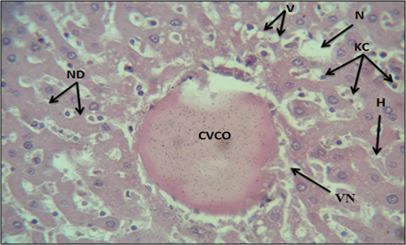
Empagliflozin is a new class of antidiabetic drugs that are specific for the sodium and glucose transporter and are taken orally. It belongs to the gliflozin family, which works to suppress blood sugar levels and also has multiple anti-inflammatory effects. Diabetes is considered one of the health problems that is increasing in its growth rate in general throughout the world. The study aims to evaluate and know the effects of some type 2 antidiabetic drugs on the histological structure of the liver of male rats in which diabetes is induced. 15 adult male rats were used in this study and they were divided into two groups. The first group was the control group, which consisted of 5 male rats. The second group included 10 male rats. This group, in turn, was divided into two secondary groups, each group containing 5 male rats. Diabetes induced by alloxan, the first secondary group remained afflicted with induced diabetes without any treatment, while the second secondary group was treated with the drug Empagliflozin at a concentration of 5 mg/kg, which was dosed to the animals for 30 days and once every day. The results obtained included congestion of the central vein, irregularity of the hepatic cells in the form of ropes, enlargement of some hepatic cells, loss of nuclei in some cells and thickening of some nuclei in others, the appearance of edema, bleeding within the sinusoids, dilatation of the sinusoids, and infiltration of inflammatory cells. The beginning of the formation of fatty tissue.
Downloads
References
[1] R. M. M. Khan, Z. J. Y. Chua, J. C. Tan, Y. Yang, Z. Liao, and Y. Zhao, "From pre-diabetes to diabetes: Diagnosis, treatments and translational research," Medicina, vol. 55, no. 9, p. 546, 2019, doi: 10.3390/medicina55090546.
[2] World Health Organization and International Diabetes Federation, Classification of Diabetes Mellitus. Geneva, Switzerland: World Health Organization, 2019.
[3] C. Faselis, A. Katsimardou, K. Imprialos, P. Deligkaris, M. Kallistratos, and K. Dimitriadis, "Microvascular complications of type 2 diabetes mellitus," Current Vascular Pharmacology, vol. 18, no. 2, pp. 117–124, 2020, doi: 10.2174/1570161117666190502103733.
[4] D. Moriconi et al., "Glomerular hyperfiltration predicts kidney function decline and mortality in type 1 and type 2 diabetes: A 21-year longitudinal study," Diabetes Care, vol. 46, no. 4, pp. 845–853, 2023, doi: 10.2337/dc22-2003.
[5] M. I. Harris, Diabetes in America. Bethesda, MD, USA: National Institutes of Health, 2013.
[6] American Diabetes Association, "Classification and diagnosis of diabetes: Standards of medical care in diabetes—2021," Diabetes Care, vol. 44, no. 1, pp. S15–S33, 2021, doi: 10.2337/dc21-S002.
[7] A. M. Ali et al., "Antidiabetic potency, antioxidant effects, and mode of actions of Citrus reticulata fruit peel hydroethanolic extract, hesperidin, and quercetin in nicotinamide/streptozotocin-induced Wistar diabetic rats," Oxidative Medicine and Cellular Longevity, vol. 2020, Art. no. 1730492, 2020, doi: 10.1155/2020/1730492.
[8] E. N. Gurzov and D. L. Eizirik, "Bcl-2 proteins in diabetes: Mitochondrial pathways of β-cell death and dysfunction," Trends in Cell Biology, vol. 21, no. 7, pp. 424–431, 2011, doi: 10.1016/j.tcb.2011.03.001.
[9] D. A. Cunha et al., "Death protein 5 and p53-upregulated modulator of apoptosis mediate endoplasmic reticulum stress–mitochondrial dialog triggering β-cell apoptosis," Diabetes, vol. 61, no. 11, pp. 2763–2775, 2012, doi: 10.2337/db12-0123.
[10] S. Steiner, "Empagliflozin, cardiovascular outcomes, and mortality in type 2 diabetes," Zeitschrift für Gefäßmedizin, vol. 13, no. 1, pp. 17–18, 2016, doi: 10.1056/NEJMoa1504720.
[11] V. Quagliariello et al., "The SGLT-2 inhibitor empagliflozin improves myocardial strain, reduces cardiac fibrosis and pro-inflammatory cytokines in non-diabetic mice treated with doxorubicin," Cardiovascular Diabetology, vol. 20, Art. no. 150, 2021, doi: 10.1186/s12933-021-01346-y.
[12] R. Saad et al., "The molecular effects of SGLT2i empagliflozin on the autophagy pathway in diabetes mellitus type 2 and its complications," Journal of Diabetes Research, vol. 2022, Art. no. 8337823, pp. 1–11, 2022, doi: 10.1155/2022/8337823.
[13] J. Cai et al., "AMPK: The key to ischemia-reperfusion injury," Journal of Cellular Physiology, vol. 237, no. 11, pp. 4079–4096, 2022, doi: 10.1002/jcp.30875.
[14] K. Houguig, S. Rkha, M. Rayadi, and N. Ouzennou, "Factors influencing therapeutic observance in diabetic subjects in the province of Essaouira (Morocco): A cross-sectional study," Ethiopian Journal of Health Sciences, vol. 32, no. 4, pp. 747–754, 2022, doi: 10.4314/ejhs.v32i4.11.
[15] A. Elksnis, M. Martinell, O. Eriksson, and D. Espes, "Heterogeneity of metabolic defects in type 2 diabetes and its relation to reactive oxygen species and alterations in beta-cell mass," Frontiers in Physiology, vol. 10, Art. no. 107, 2019, doi: 10.3389/fphys.2019.00107.
[16] E. Sokolowska and A. Blachnio-Zabielska, "The role of ceramides in insulin resistance," Frontiers in Endocrinology, vol. 10, Art. no. 577, 2019, doi: 10.3389/fendo.2019.00577.
[17] Z. A. Jigheh et al., "Empagliflozin alleviates renal inflammation and oxidative stress in streptozotocin-induced diabetic rats partly by repressing HMGB1-TLR4 receptor axis," Iranian Journal of Basic Medical Sciences, vol. 22, no. 4, pp. 384–391, 2019, doi: 10.22038/ijbms.2019.31788.7651.
[18] I. A. Okafor, U. S. Nnamah, and J. Nnaka, "Liver function analyses and spleen histology assessment following the co-administration of cisplatin and methanolic extract of Portulaca oleracea in Wistar rats," Natural Product Sciences, vol. 26, no. 3, pp. 252–258, 2020, doi: 10.20307/nps.2020.26.3.252.
[19] E. Ferrari and M. Rasponi, "Liver–heart on chip models for drug safety," APL Bioengineering, vol. 5, no. 3, Art. no. 031504, 2021, doi: 10.1063/5.0048986.
[20] K. Reyes-Gordillo, R. Shah, and P. Muriel, "Oxidative stress and inflammation in hepatic diseases: Current and future therapy," Oxidative Medicine and Cellular Longevity, vol. 2017, Art. no. 3140673, 2017, doi: 10.1155/2017/3140673.
[21] N. Sandhu and V. Navarro, "Drug-induced liver injury in GI practice," Hepatology Communications, vol. 4, no. 5, pp. 631–645, 2020, doi: 10.1002/hep4.1503.
[22] C. Ronco and L. Di Lullo, "Cardiorenal syndrome," Heart Failure Clinics, vol. 10, no. 2, pp. 251–280, 2014.
Downloads
Published
Issue
Section
License
Copyright (c) 2026 Iraqi Journal for Applied Science

This work is licensed under a Creative Commons Attribution 4.0 International License.
Licenses and Copyright
The copyright of this article is retained by the author(s).
This article is published by the Iraqi Journal for Applied Science (IJAS) under the Creative Commons Attribution 4.0 International License (CC BY 4.0).
This license permits unrestricted use, distribution, reproduction, adaptation, and reuse of the work in any medium or format, including commercial use, provided that appropriate credit is given to the original author(s) and the source, a link to the license is provided, and any modifications are indicated.
Authors retain full copyright of their work and grant IJAS a non-exclusive license to publish, archive, preserve, and disseminate the article, including registration of metadata and Digital Object Identifiers (DOIs) through Crossref.
Any third-party material included in this article remains subject to its respective copyright and licensing conditions. Authors are responsible for obtaining all necessary permissions for such material before publication.
For complete details regarding copyright, licensing, permissions, and reuse, please refer to the journal's Copyright and Licensing Policy available on the IJAS website.
License: https://creativecommons.org/licenses/by/4.0/
How to Cite