Robust Patient-Level Evaluation of Post-Training Quantization for Edge-Deployable MPox Skin Lesion Classification
DOI:
https://doi.org/10.69923/qfnn3n63Keywords:
MPox classification, Patient-level split, INT8 inference, Post-training quantization, Medical imageAbstract
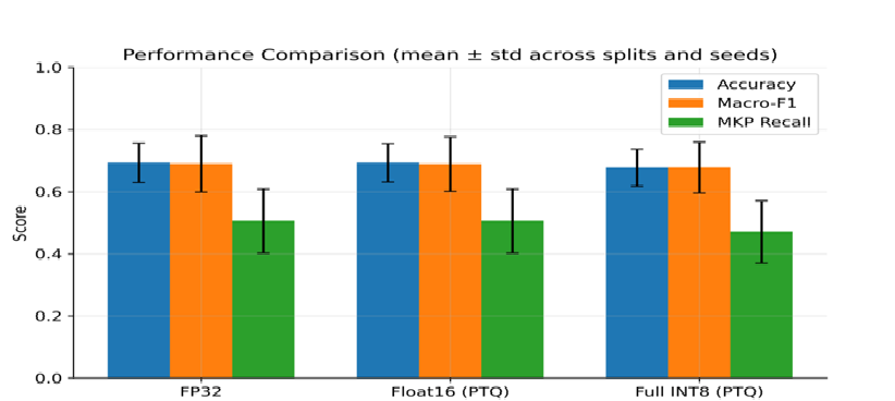
In this study, we implemented Post-Training Quantization (PTQ) for Mpox skin lesion classification using a patient-level evaluation protocol based on a stringent pre-registered experimental design. The publicly available MSLD v2.0 dataset was preprocessed at the pixel level, resulting in 755 unique original images from six diagnostic classes representing 524 patients. A stratified patient-level split (80% training, 10% validation, and 10% testing) was applied to prevent data leakage. Each experiment was repeated across three independent data splits and three training seeds to assess performance variability and robustness. A two-stage transfer learning framework based on MobileNetV2 was trained and evaluated using FP32, Float16 PTQ, and full INT8 quantization implemented through TensorFlow Lite (TFLite). The FP32 baseline achieved an accuracy of 69%, a macro F1-score of 69%, and a macro-AUC of 94.7%, with a model size of 4.27 MB. Float16 quantization maintained comparable predictive performance and model size, whereas full INT8 quantization reduced the model size to 2.59 MB, representing a 39% reduction compared with the FP32 baseline. Top-2 accuracy remained stable at approximately 89% across all precision regimes, supporting the feasibility of deployment in triage-oriented clinical settings. Variability analyses revealed that performance fluctuations were driven primarily by patient partitioning rather than reduced numerical precision, demonstrating that INT8 quantization preserves clinically relevant performance while substantially reducing computational and storage requirements for deployment in resource-constrained edge environments.
Downloads
References
[1] A. H. Thieme et al., “A deep-learning algorithm to classify skin lesions from mpox virus infection,” Nat. Med., vol. 29, no. 3, pp. 738–747, 2023. DOI: 10.1038/s41591-023-02225-7
[2] S. N. Ali et al., “A web-based mpox skin lesion detection system using state-of-the-art deep learning models considering racial diversity,” Biomed. Signal Process. Control, vol. 98, p. 106742, 2024. DOI: 10.1016/j.bspc.2024.106742
[3] A. S. Jaradat et al., “Automated monkeypox skin lesion detection using deep learning and transfer learning techniques,” Int. J. Environ. Res. Public Health, vol. 20, no. 5, p. 4422, 2023. DOI: 10.3390/ijerph20054422
[4] E. F. Rivera-Guzmán, L. F. Guerrero-Vásquez, and V. E. Robles-Bykbaev, “Quantization of Deep Neural Networks for Medical Image Analysis: A Systematic Review and Meta-Analysis,” Technologies, vol. 14, no. 1, p. 76, 2026. DOI: 10.3390/technologies14010076
[5] C. Qu et al., “Post-Training Quantization for 3D Medical Image Segmentation: A Practical Study on Real Inference Engines,” arXiv Prepr. arXiv2501.17343, 2025. DOI: 10.48550/arXiv.2501.17343
[6] M. S. Elhadidy, A. T. Elgohr, A. Mousa, A. Safwat, R. I. Abdelfatah, and H. M. Kasem, “Benchmarking pre-trained CNNs and vision transformers for Mpox-related dermatological image classification on MSLD v2. 0,” Results Eng., p. 108071, 2025. DOI: 10.1016/j.rineng.2025.108071
[7] H. Alghoraibi et al., “Deep Learning-Based Mpox Skin Lesion Detection and Real-Time Monitoring in a Smart Healthcare System,” Diagnostics, vol. 15, no. 19, p. 2505, 2025. DOI: 10.3390/diagnostics15192505
[8] T. Singh and A. Solanki, “ResEffNet: Web-Based Mpox Skin Lesion Detection System,” in International Conference on Data Analytics & Management, 2025, pp. 314–325. DOI: 10.1007/978-3-032-03740-4_25
[9] G. Yolcu Oztel, “Vision transformer and CNN-based skin lesion analysis: classification of monkeypox,” Multimed. Tools Appl., vol. 83, no. 28, pp. 71909–71923, 2024. DOI: 10.1007/s11042-024-19757-w
[10] M. A. Rahim, M. N. Arefin, M. M. Rahman, M. A. Hossain, and A. Moustafa, “An empirical study for the early detection of Mpox from skin lesion images using pretrained CNN models leveraging XAI technique,”. DOI: 10.48550/arXiv.2507.15915
[11] X. Cao, W. Ye, K. Moise, and M. Coffee, “Mpoxvlm: A vision-language model for diagnosing skin lesions from mpox virus infection,” arXiv Prepr. arXiv2411.10888, 2024. DOI: 10.48550/arXiv.2411.10888
[12] M. Sandler, A. Howard, M. Zhu, A. Zhmoginov, and L.-C. Chen, “Mobilenetv2: Inverted residuals and linear bottlenecks,” in Proceedings of the IEEE conference on computer vision and pattern recognition, 2018, pp. 4510–4520. DOI: 10.1109/CVPR.2018.00474
[13] S. Vuran, M. Ucan, M. Akin, and M. Kaya, “Multi-classification of skin lesion images including Mpox disease using transformer-based deep learning architectures,” Diagnostics, vol. 15, no. 3, p. 374, 2025. DOI: 10.3390/diagnostics15030374
Downloads
Published
Issue
Section
License
Copyright (c) 2026 Iraqi Journal for Applied Science

This work is licensed under a Creative Commons Attribution 4.0 International License.
Licenses and Copyright
The copyright of this article is retained by the author(s).
This article is published by the Iraqi Journal for Applied Science (IJAS) under the Creative Commons Attribution 4.0 International License (CC BY 4.0).
This license permits unrestricted use, distribution, reproduction, adaptation, and reuse of the work in any medium or format, including commercial use, provided that appropriate credit is given to the original author(s) and the source, a link to the license is provided, and any modifications are indicated.
Authors retain full copyright of their work and grant IJAS a non-exclusive license to publish, archive, preserve, and disseminate the article, including registration of metadata and Digital Object Identifiers (DOIs) through Crossref.
Any third-party material included in this article remains subject to its respective copyright and licensing conditions. Authors are responsible for obtaining all necessary permissions for such material before publication.
For complete details regarding copyright, licensing, permissions, and reuse, please refer to the journal's Copyright and Licensing Policy available on the IJAS website.
License: https://creativecommons.org/licenses/by/4.0/
How to Cite