Green Synthesis by Berberine and Biological Evaluation of Fe₂O₃ and FeS Nanoparticles
DOI:
https://doi.org/10.69923/n8gabj57Keywords:
Green synthesis; Berberine extract; Fe2O3 nanoparticles; FeS nanoparticles; Resazurin MIC; Antioxidant activity; Biosafety.Abstract
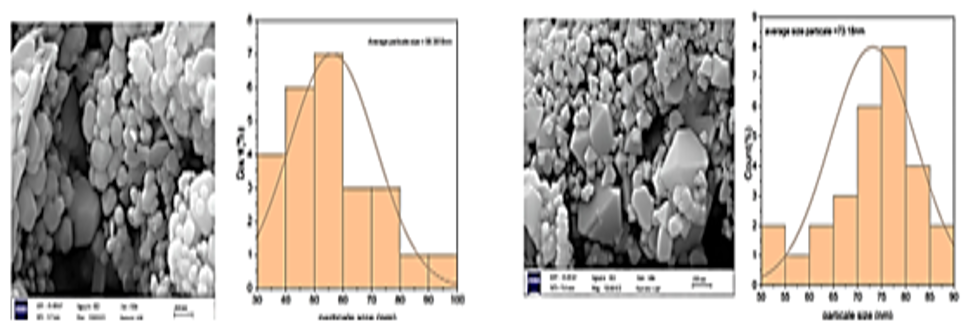
This study demonstrates a green and cost-effective method for fabricating crystalline hematite Fe₂O₃ and iron sulfide FeS nanoparticles using a bioactive alkaline berberine extract. The berberine-coated nanoparticles exhibited high structural integrity and excellent colloidal stability, supported by strong negative zeta potentials of -28.2 mV for Fe₂O₃ and -30.9 mV for FeS. Biologically, both nanostructures demonstrated potent concentration-dependent antimicrobial activity against S. aureus and Escherichia coli: the Fe₂O₃ nanoparticles specifically targeted S. aureus at a minimum inhibitory concentration (MIC) of 32 µg/ml (a concentration below the MIC of 16 µg/ml). FeS NPs exhibited balanced activity against both strains at a minimum inhibitory concentration (MIC) of 64 µg/ml (lower MIC: 32 µg/ml). Furthermore, ferric oxide NPs demonstrated superior antioxidant capacity as measured by DPPH (88.4% inhibition vs. 60.5% for ferric sulfide). Importantly, in vitro assays confirmed minimal cytotoxicity (0.52% for ferric oxide and 0.89% for ferric sulfide) at therapeutic concentrations, making them promising platforms for biomedical applications.
Downloads
References
[1] A. Khan et al., “Biogenic nanoparticles and green nanocomposites as sustainable antimicrobial strategies against bacteria, fungi and viruses,” Nano-Horizons: Journal of Nanosciences and Nanotechnologies, vol. 5, Feb. 2026, doi: 10.25159/3005-2602/20247.
[2] A. Ali et al., “Synthesis, characterization, applications, and challenges of iron oxide nanoparticles,” Nanotechnol. Sci. Appl., vol. 9, pp. 49–67, Aug. 2016, doi: 10.2147/nsa. s99986.
[3] L. Arias, J. Pessan, A. Vieira, T. Lima, A. Delbem, and D. Monteiro, “Iron Oxide Nanoparticles for Biomedical Applications: A Perspective on Synthesis, Drugs, Antimicrobial Activity, and Toxicity,” Antibiotics, vol. 7, no. 2, p. 46, June 2018, doi: 10.3390/antibiotics7020046.
[4] J. Singh, T. Dutta, K.-H. Kim, M. Rawat, P. Samddar, and P. Kumar, “‘Green’ synthesis of metals and their oxide nanoparticles: applications for environmental remediation,” J. Nanobiotechnol., vol. 16, no. 1, Oct. 2018, doi: 10.1186/s12951-018-0408-4.
[5] M. S. Akhtar, J. Panwar, and Y.-S. Yun, “Biogenic synthesis of metallic nanoparticles by plant extracts,” ACS Sustain. Chem. Eng., vol. 1, no. 6, pp. 591–602, Apr. 2013, doi: 10.1021/sc300118u.
[6] Neag, M. A., et al., “Berberine: Botanical occurrence, traditional uses, extraction methods, and relevance in cardiovascular, metabolic, hepatic, and Renal Disorders,” Frontiers in Pharmacology, vol. 9, no. 557, Aug. 2018, doi: 10.3389/fphar.2018.00557.
[7] C. Marques, L. Grenho, M. H. Fernandes, and S. A. Costa Lima, “Improving the antimicrobial potency of Berberine for Endodontic Canal Irrigation Using Polymeric Nanoparticles,” Pharmaceutics, vol. 16, no. 6, p. 786, June 2024, doi: 10.3390/pharmaceutics16060786.
[8] W. G. Pearl et al., “Berberine mediated fluorescent gold nanoclusters in biomimetic erythrocyte ghosts as a nanocarrier for enhanced photodynamic treatment,” RSC Adv., vol. 14, no. 5, pp. 3321–3334, 2024, doi: 10.1039/d3ra08299g.
[9] S. Ezzine, H. Ferjani, O. E. Ogunjinmi, and D. C. Onwudiwe, “Hematite nanoparticles synthesized by green route: characterization, anticancer and antioxidant activities,” Inorganics, vol. 13, no. 5, p. 167, May 2025, doi: 10.3390/inorganics13050167.
[10] S. M. B. Bataineh et al., “Synergistic effect of magnetic iron oxide nanoparticles with medicinal plant extracts against resistant bacterial strains,” Magnetochemistry, vol. 10, no. 7, p. 49, July 2024, doi: 10.3390/magnetochemistry10070049.
[11] N. P. S. Chauhan et al., “Recent advances in the green synthesis of metal and metal oxide nanoparticles from plant extracts for biomedical and water remediation applications,” N. Nanotechnol., vol. 9, p. 100451, June 2026, https://doi.org/10.1016/j.nxnano.2026.100451.
[12] Jarullah, A. A., Hammadi, M., & Hassan, “Green Synthesis of Nano Zn3(PO4)2 Using Curcuma Extract and Their Effect on SK-OV-3 Human Ovarian Cancer Cell Lines,” Biological Sciences - PJSIR, vol. 68, no. 2, pp. 166–171, July 2025, doi: 10.52763/pjsir.biol.sci.68.2.2025.166.171.
[13] L. G. Calvo, S. Corral-Orbe, R.-A. Villarino, S. Sánchez, and T. de Miguel, “Employment of a Resazurin Viability-Based Assay for Minimum Inhibitory and Bactericidal Concentration Determination,” Pharmaceuticals, vol. 19, no. 3, p. 505, Mar. 2026, doi: 10.3390/ph19030505.
[14] J. A. A. Abdullah, L. Salah Eddine, B. Abderrhmane, M. Alonso-González, A. Guerrero, and A. Romero, “Green synthesis and characterization of iron oxide nanoparticles by pheonix dactylifera leaf extract and evaluation of their antioxidant activity,” Sustainable Chemistry and Pharmacy, vol. 17, p. 100280, Sept. 2020, doi: 10.1016/j.scp.2020.100280.
[15] S. Saif, A. Tahir, and Y. Chen, “Green Synthesis of Iron Nanoparticles and Their Environmental Applications and Implications,” Nanomaterials, vol. 6, no. 11, p. 209, Nov. 2016, doi: 10.3390/nano6110209
[16] S. Marimuthu, P. Kannan, and G. Maduraiveeran, “Transition metal sulfide nanostructures: synthesis and application in metal-air batteries,” Nano Express, vol. 5, no. 2, p. 022005, June 2024, doi: 10.1088/2632-959x/ad585c.
[17] Nagaraj, A. et al., “Fe2O3-type iron oxide nanoparticles from Aerva lanata leaf extract exhibit antibiofilm, discriminatory toxicity in cancer cells, and theranostic against oxidative stress in zebrafish,” Chem. Phys. Impact, vol. 10, p. 100849, June 2025, doi: 10.1016/j.chphi.2025.100849.
[18] Z. U. H. Khan et al., “Biomedical applications of green synthesized Nobel metal nanoparticles,” Journal of Photochemistry and Photobiology B: Biology, vol. 173, pp. 150–164, Aug. 2017, doi: 10.1016/j.jphotobiol.2017.05.034.
[19] “Synergistic antioxidant activity of green-synthesized iron oxide nanoparticles: a comparative study of Capparis decidua phytochemical-mediated synthesis and in vitro evaluation,” J Chem Health Risks, Apr. 2026, doi: 10.52783/jchr.v16.i2.13045.
[20] Y. El Mendili et al., “Magnetic interactions in γ-Fe2O3@SiO2 nanocomposites,” J. Appl. Phys., vol. 116, no. 5, Aug. 2014, doi: 10.1063/1.4891497.
[21] I. T. Sines, D. D. Vaughn II, R. Misra, E. J. Popczun, and R. E. Schaak, “Synthesis of tetragonal mackinawite-type FeS nanosheets by solvothermal crystallization,” J. Solid State Chem., vol. 196, pp. 17–20, Dec. 2012, doi: 10.1016/j.jssc.2012.07.056.
[22] A. SAAD and M. ELHADI, “ESTIMATION OF CRYSTALLITE SIZE, LATTICE PARAMETER, INTERNAL STRAIN AND CRYSTAL IMPURIFICATION OF NANOCRYSTALLINE Al3Ni20Bx ALLOY BY WILLIAMSON-HALL METHOD,” Journal of Ovonic Research, vol. 16, no. 5, pp. 323–335, Sept. 2020, doi: 10.15251/jor.2020.165.323.
[23] N. M. Noah and P. M. Ndangili, “Green synthesis of nanomaterials from sustainable materials for biosensors and drug delivery,” Sensors International, 3, 100166. https://doi.org/10.1016/j.sintl.2022.100166.
[24] Y.-C. Lin, Z.-R. Lin, L.-W. Tsai, E. Perevedentseva, A. Karmenyan, and C.-L. Cheng, “Spectral analysis of Nanodiamond-berberine complex interaction with living cells for nanoparticle mediated drug delivery,” J. Biomed. Photonics Eng., vol. 3, no. 1, p. 010305, 2017, https://doi.org/10.18287/jbpe17.03.010305.
[25] A. Shafiq, A. R. Deshmukh, K. AbouAitah, and B.-S. Kim, “Green synthesis of controlled shape silver nanostructures and their peroxidase, catalytic degradation, and antibacterial activity,” J. Funct. Biomater., vol. 14, no. 6, p. 325, June 2023, doi: 10.3390/jfb14060325.
[26] Y. Zhao et al., “Self-assembled Berberine-Sinapic Acid Nanomedicine for Synergistic Chemotherapy,” Molecules, vol. 31, no. 4, p. 621, Feb. 2026, doi: 10.3390/molecules31040621.
[27] J. Shi, Z. Ai, and L. Zhang, “Fe@Fe2O3 core-shell nanowires enhanced Fenton oxidation by accelerating the Fe(III)/Fe(II) cycles,” Water Research, 59, 145–153. https://doi.org/10.1016/j.watres.2014.04.015.
[28] V. Poderys et al., “Upconverting nanoparticles functionalized with protein–gold nanoclusters and chlorin e6 for near-infrared-activated photodynamic therapy,” Nanomaterials, vol. 16, no. 7, p. 417, Mar. 2026, doi: 10.3390/nano16070417.
[29] Resazurin Microtiter Assay Plate method for detection of susceptibility of multidrug resistant Mycobacterium tuberculosis to second-line anti-tuberculous drugs,” Eur J Chest Dis Tuberc, vol. 62, no. 2, pp. 241–247, Apr. 2013, doi: 10.1016/j.ejcdt.2013.05.008.
[30] T. D. Sallal and A. M. Abbas, “Preparation and characterization of nano-iron oxide by using Iraqi Orange Plant Extract and Testing for Adsorption Efficiency,” International Journal of Biology and Chemistry, 18(1). https://doi.org/10.26577/ijbch202518112.
Downloads
Published
License
Copyright (c) 2026 Fatima Hamdi (Author)

This work is licensed under a Creative Commons Attribution 4.0 International License.
Licenses and Copyright
The copyright of this article is retained by the author(s).
This article is published by the Iraqi Journal for Applied Science (IJAS) under the Creative Commons Attribution 4.0 International License (CC BY 4.0).
This license permits unrestricted use, distribution, reproduction, adaptation, and reuse of the work in any medium or format, including commercial use, provided that appropriate credit is given to the original author(s) and the source, a link to the license is provided, and any modifications are indicated.
Authors retain full copyright of their work and grant IJAS a non-exclusive license to publish, archive, preserve, and disseminate the article, including registration of metadata and Digital Object Identifiers (DOIs) through Crossref.
Any third-party material included in this article remains subject to its respective copyright and licensing conditions. Authors are responsible for obtaining all necessary permissions for such material before publication.
For complete details regarding copyright, licensing, permissions, and reuse, please refer to the journal's Copyright and Licensing Policy available on the IJAS website.
License: https://creativecommons.org/licenses/by/4.0/
How to Cite